Sensibilité accrue et plage de détection élargie grâce à de nouveaux systèmes de détection
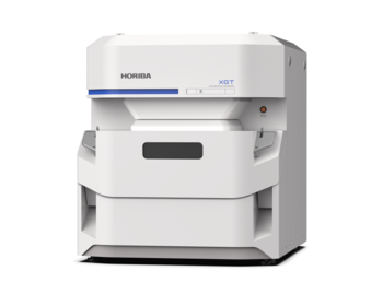
De nouveaux systèmes de détection avancés permettent au XGT-9000 Pro d'atteindre un niveau de sensibilité inédit et au XGT-9000 Expert d'afficher une sensibilité ultra-haute performance et la gamme d'éléments détectables la plus large. L'amélioration de la sensibilité réduit le temps de mesure et renforce l'efficacité, tandis que l'enrichissement de la gamme d'éléments détectables élargit les possibilités d'application.
(Gauche) Comparaison de l'intensité du Cu. (Droite) Comparaison de l'intensité des éléments super légers.
[1][2] Tous les résultats sont comparés entre le XGT-9000 Pro/Expert et les modèles micro-XRF classiques d'��������ֱ��
Imagerie macro et micro de haute qualité avec sélection de sondes multiples
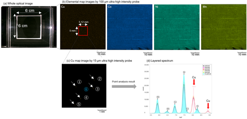
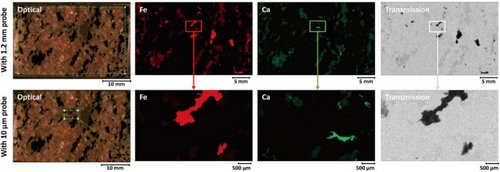
La série XGT-9000 offre des performances et une flexibilité optimales grâce à son système d'excitation bien conçu, comprenant un générateur de rayons X à haut rendement (jusqu'à 50 kV et 1 000 µA) et un large choix de tailles de point de sonde jusqu'à 10 microns et 1,2 mm. Il est possible d'installer plusieurs sondes dans l'instrument et de sélectionner dans le logiciel celles que vous souhaitez utiliser. Ces instruments permettent une imagerie macro et micro rapide et de haute qualité qui ne compromet en rien les besoins de résolution spatiale et de temps de mesure sur une zone cartographique étendue. Deux sondes ultra haute intensité de 15 µm et 100 µm sont disponibles.
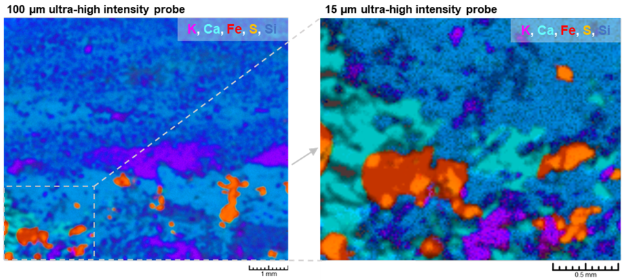
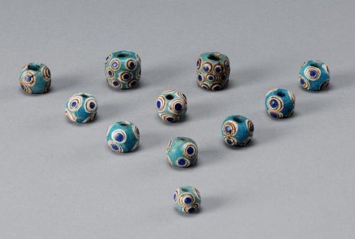
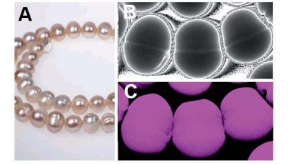
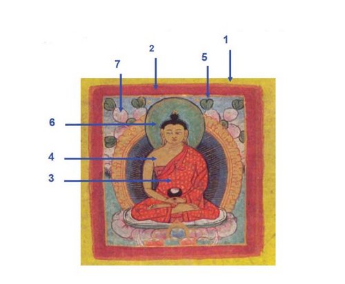
Images élémentaires en couches sur une pierre de Lapis-lazuli avec plusieurs sondes
(Gauche) Balayage rapide avec une sonde ultra haute intensité de 100 µm. (À droite) Imagerie détaillée avec une sonde ultra haute intensité de 15 µm.
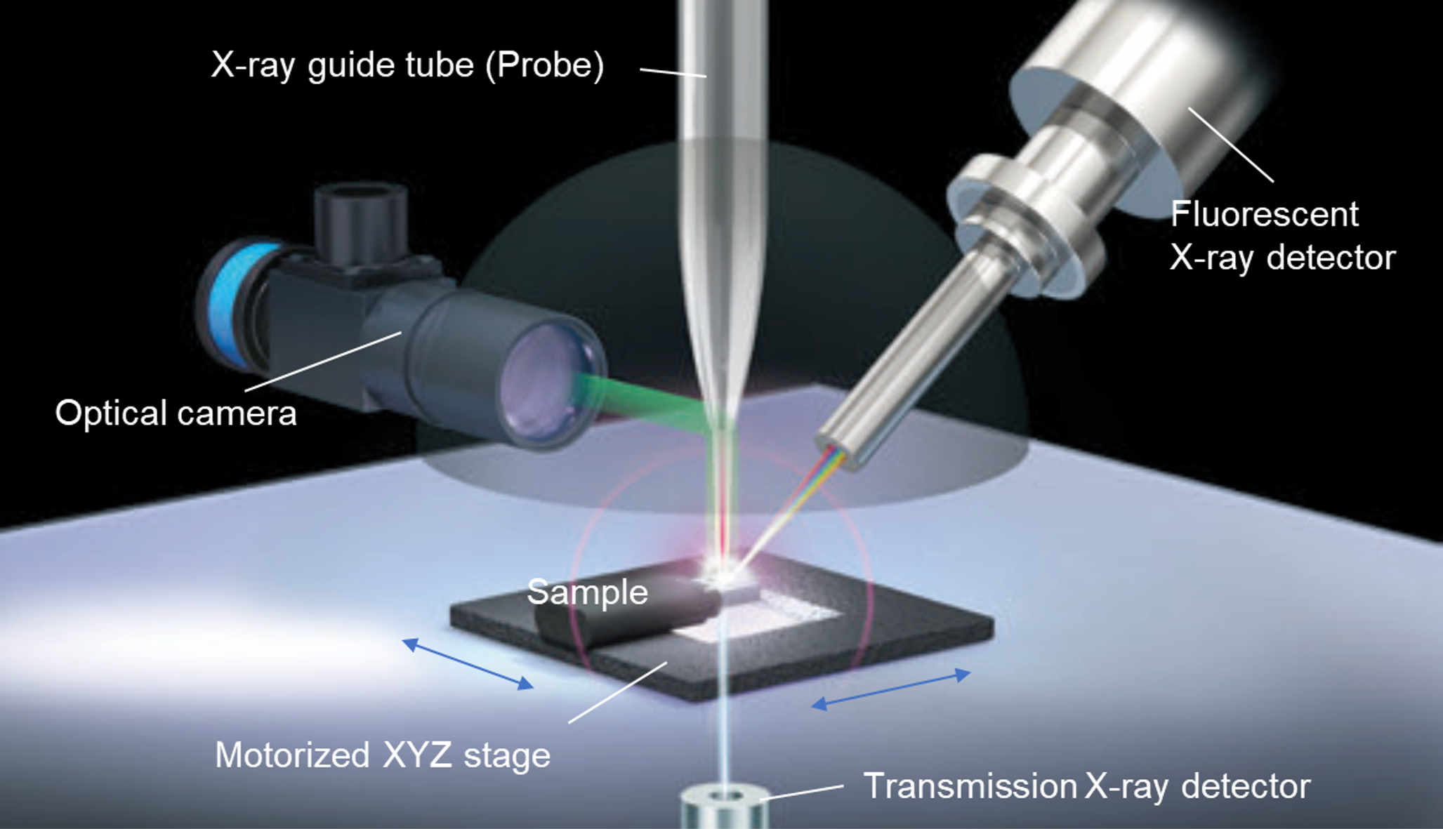
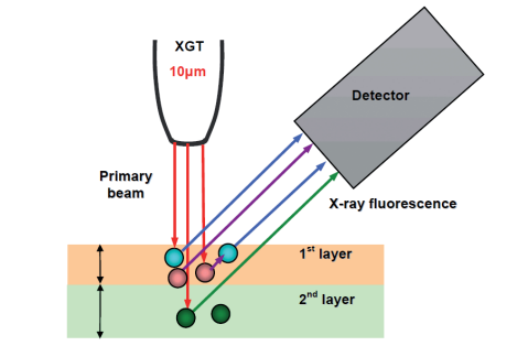
Deux types de détecteurs pour les rayons X fluorescents et les rayons X émis
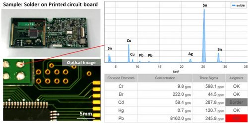
La série XGT-9000 propose deux types de détecteurs : un détecteur de rayons X fluorescents qui indique la distribution élémentaire d'un échantillon et un détecteur de rayons X émis qui indique la structure interne d'un échantillon. Le XGT-9000 peut obtenir simultanément les deux types d'images de la même zone, ce qui est particulièrement utile pour mieux comprendre l'électronique, les pierres précieuses (y compris les inclusions) et les échantillons biologiques.
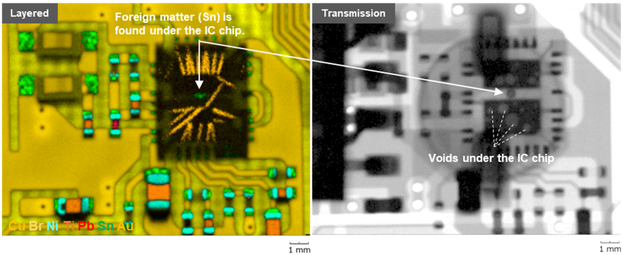
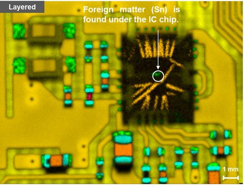
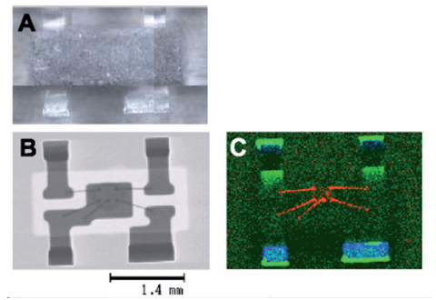
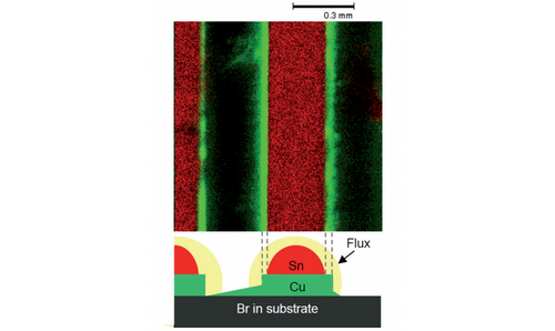
Imagerie simultanée d'une image élémentaire et d'une image des rayons X émis d'un circuit imprimé
(Gauche) L'image élémentaire en couches a révélé la présence d'un corps étranger sous la puce électronique. (Droite) L'image des rayons X émis a révélé la présence de nombreux vides sous la puce.
Images optiques haute résolution et brillantes
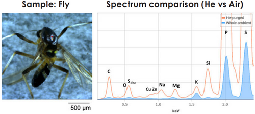
L'obtention d'images claires est essentielle pour l'analyse micro-XRF. La série XGT-9000 propose des caméras haute résolution pour saisir l'image de l'échantillon complet et de ses ��é�ٲ������� jusqu'au niveau microscopique. Il est possible d'agrandir ou de réduire numériquement les deux images pour identifier librement la meilleure position à analyser sur la surface de l'échantillon. En outre, grâce aux nombreux systèmes d'éclairage, vous pouvez obtenir des images optiques brillantes de votre échantillon cible.
(Gauche) Image globale (Droite) Image de détail
Image optique haute résolution et brillante d'une mouche
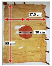
Nombreuses applications de la chambre à échantillon
La série XGT-9000 permet de traiter une grande variété d'échantillons tels que des fragments de taille micro, des circuits imprimés, des pièces de monnaie, des feuilles, des poudres, des liquides et des plaquettes. Différents types de porte-échantillon peuvent être fournis. Il est également possible de sélectionner jusqu'à 4 environnements de mesure pour augmenter la sensibilité.
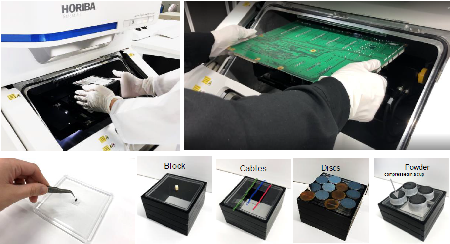
Nombreuses applications de la chambre à échantillon
Logiciel flexible et convivial
Le logiciel du XGT-9000 est doté d'une interface très flexible et conviviale, qui permet de visualiser en un coup d'œil l'arborescence des données, les images optiques, les résultats du spectre, le tableau périodique et les résultats de l'imagerie cartographique. Et grâce à sa flexibilité, les utilisateurs peuvent personnaliser la disposition de l'écran. Sans oublier que les résultats peuvent être affichés sur plusieurs écrans, ce qui permet d'assurer une meilleure visibilité des résultats et d'améliorer l'analyse des données.
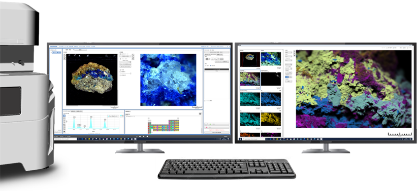
Exemple de présentation du logiciel sur deux écrans
Il est possible d'ajouter des modules avancés aux fonctions standard de la suite logicielle pour une expérience utilisateur plus complète.
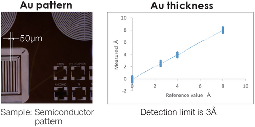
- Module FPM multicouche pour la mesure de l'épaisseur avec/sans échantillons standard
- Module RoHS pour le criblage RoHS
- Module de file d'attente pour les mesures multiples automatisées en mode sans surveillance
- Module de recherche de particules pour l'analyse des particules et l'analyse colocalisée
- Module LabSpec Link pour le transfert des données vers LabSpec 6 à des fins d'analyse multivariée
Publications
Exemples d'applications (en anglais)
Vidéo de présentation du produit
Dispositif de transfert pour l'analyse d'échantillons sensibles à l'air (accessoire en option)